- Disclaimer / Privacy
© 2025 All Rights Reserved.
Danbury Office
51-53 Kenosia Avenue
Danbury, CT 06810
203.748.0330
New Milford Office
120 Park Lane
Suite A202
New Milford, CT 06776
203.748.0330

Kidney stones may be as small as a grain of sand or larger than a golf ball. They may be smooth, round, jagged, spiky, or asymmetrical, depending on their composition. Most stones are yellow or brown in color, but variations in chemical composition can produce stones that are tan, gold, or black.
Some stones stay in the kidney and produce no symptoms. Other stones break loose and travel down the urinary tract. The smallest, smoothest stones may pass out of the body with little resistance, and the patient may experience minimal discomfort. Large, irregularly shaped stones can become lodged in a ureter (tube that carries urine from the kidney to the bladder), the bladder, or the urethra (tube that carries urine from the bladder out of the body) and cause intense pain.
A lodged stone can block the flow of urine, causing waste and pressure to build up in the kidneys. Such a condition must be corrected swiftly to prevent serious kidney damage and other medical problems. The stone can be removed surgically, or can be broken up and then passed naturally out of the body.
Men are four to five times more likely to develop the disease than women. An estimated 10% of men and 5% of women between the ages of 30 to 50 will suffer from kidney stones. Kidney stones are four to five times more common in Caucasians than in African Americans. Most people who develop kidney stones experience their first episode between the ages of 20 and 30. Kidney stone disease usually continues throughout life, particularly in men.
A stone may grow until it begins to block the flow of urine. Patients often endure months, even years, of chronic back pain without associating their discomfort with a kidney problem.
Sometimes a small stone will pass without producing any symptoms, or the symptoms are so mild that the patient attributes them to backache, muscle strain, or the onset of a flu-type illness. Such patients are considered asymptomatic and often experience recurrent urinary tract infections before the cause is accurately diagnosed.
Classic symptoms associated with a lodged stone that causes urinary tract irritation and/or obstructs the flow of urine are more common and the symptoms are immediate and severe. This condition is known as acute ureteral colic.
Acute ureteral colic comes on suddenly, often at night or early in the morning. It may feel like an attack of appendicitis, gastroenteritis, or colitis. Pain usually starts in the back, at the waist, or in the flank, stomach or groin. (“Teeth-gritting agony” is the way one expert characterizes it.) The pain can radiate down the leg or into a man’s testes or tip of the penis, depending on where the stone has lodged.
Nausea, vomiting, chills, fever, and elevated blood pressure are common, and despite the discomfort, the patient often cannot sit or lie still. There may be an urgent need to urinate frequently, accompanied by a burning sensation during urination.
Hematuria (blood in the urine) is another classic symptom. The blood may be clearly visible, or it may be seen only through microscopic examination. Often, the patient’s urine is unusually dark and/or cloudy and has a strong odor.
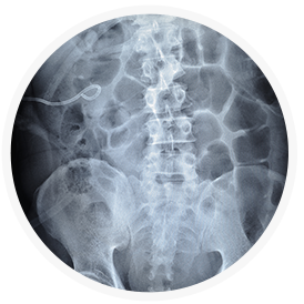
![]()
Ureteroscopy is the technique of choice with smaller stones that lodge in the mid and lower sections of the ureter. A ureteroscope (flexible, fiberoptic instrument resembling a long, thin telescope) is inserted through the urethra and bladder to the stone. The urologist can locate the stone visually and remove it with a small basket-like device inserted through the ureteroscope. This procedure is called endoscopic basket extraction. A laser may be used to break the stone into smaller pieces, which can be passed by the patient. Ureteroscopy is performed under general or local anesthesia on an outpatient basis. The urologist often places a small silicone tube (stent) into the ureter to relieve swelling and facilitate healing.
A variety of nonsurgical techniques have been developed to crush or pulverize kidney stones. Lithotripsy uses a machine called a lithotripter to project shock waves or sonic pulses against the stone and break it into tiny particles that can pass naturally in the patient’s urine. This can be done in several ways, depending on the size and location of the stone.
Patients undergoing lithotripsy are given a sedative and a general or local anesthetic. Shock waves are focused on the kidney stone at a rate of approximately one per second and the therapy may last over an hour. Bruising may result from the shock waves and discomfort may be experienced as the crushed calculi are passed; but, most patients resume normal activity in a few days.
Lithotripsy is highly effective for stones in the kidney and upper ureter. More than one treatment may be required. Rarely, a catheter may be inserted through a small incision in the back to drain the kidney and remove the stone fragments. Patients with very large stones or complicating medical conditions may require different treatment.
Also known as (Percutaneous Nephrolithotomy, or PCNL). Some kidney stones cannot be treated by ESWL or Ureteroscopy (see above) for a variety of reasons. Stones that are large (>2cm), impacted stones of the kidney or ureter, or “staghorn” stones of the kidney often cannot be treated with less invasive procedures. A PCNL involves two steps – usually done the same day. The first is the placement of a nephrostomy tube under light anesthesia by the intervention radiologist in the radiology suite. A nephrostomy tube is a thin (3mm) tube that enters the kidney directly through the skin in the back or flank. The second step is the actual breaking and removal of the stones (nephrolithotomy) under general anesthesia by the urological surgeon in the operating room. The PCNL has a very high success rate of removing all the stones in one procedure. Post-operatively, a nephrostomy tube is left in place (in the kidney) in addition to a Foley catheter (in the bladder). These are usually removed within the first 48 hours and the patient is usually discharged from the hospital in 1-2 days.